


From our childhood we have been used to running, jumping, boys love to climb and play football, ropes girls and more.And the active lifestyle has been so entering the human mind that over the years when the muscle is pulling somewhere, somewhere the joint becomes ill, the person does not even pay attention-"Well, think how many times it hurts."Here, in today's article, we will also talk why the knee can also hurt whether this is always the usual result of sharp movement.
What is arthrosis?
Arthrosis -a group of diseases of the musculoskeletal system of different origin, but with similar biological, morphological and clinical manifestations.The basis of their development is the degenerative lesion of all components of the joint, mainly cartilage, thin bone, synovial membrane, ligaments, capsules and periarticular muscles, with the formation of marginal osteophytes and clear or hidden moderately pronounced synovitis.Because with this disease, pathological changes capture both cartilage and bone tissue.
Arthrosis is often called Osteoarthrosisand sometimes OsteoarthritisS
Statistics (Epidemiology)
Among all joint diseases, arthrosis is up to 80% of cases.
The disease develops mainly in middle and old age.At a young age, arthrosis can occur after joint injuries, inflammatory processes, and with congenital pathology of the musculoskeletal system.
X -ray signs of arthrosis are found in most people over 65 and almost 95%for 70 years.
Women suffer from arthrosis almost twice as often as men.The degree of morbidity increases during postmenopause.
A major role in the development of arthrosis is played by hereditary factors.The incidence of the disease in families of patients with osteoarthritis has been found to be twice as high as in the population as a whole, and the development of arthrosis in people with congenital defects in the musculoskeletal system increases by 7-8 times.
Arthrosis - ICD
- MKB-10: M15-M19, M47
- MKB-9: 715
- MKB-9-KM: 715.3
Symptoms of arthrosis (clinical picture)
The clinical manifestations of the disease and their severity depends on the location of the pathological process, the state of the patient's health and the image of his life.
The first signs of arthrosis
Arthrosis often begins gradually, imperceptible to the patient.
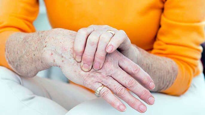
The first symptom of the disease is usually short -term easy pain in the joints (arthralgia), which carries the greatest workload.These are, first of all, the joints of the lower limbs-the pine, the hip joint, the plus-balax joints on the first thumb of the foot.From the joints of the upper limb, more often the interfal joints, the joint joint of the carpal of the thumb of the brush are more commonly affected.
Arthrosis usually begins with the lesion of one joint, but after a while other joints are involved in the process.
The main symptoms of arthrosis
With arthrosis, patients complain of pain, crunching, restriction of joint movements, swelling and joint deformity.
Separately, it is worth opting for the nature of the pain.With arthrosis, mechanical and initial pain are possible.Mechanical pain occurs when loaded on the affected joint.Such pain is worried mainly in the evening at rest, disappears after a few hours of rest.The appearance of this type of pain is associated with a gradual increase in bone pressure during exercise.Pressure causes bone rays and irritation of painful bone tissue.
Starting pain occurs at the beginning of the walk, then quickly stops and reappears during exercise.Starting pain can occur by rubbing the joint surfaces of the affected joint.Small particles of necrotic cartilage fall on cartilage surfaces.In the first steps, these particles are pushed into the cavity of the joint bag and the pain ceases.
With arthrosis, pain can be associated with periarthritis and trend (inflammation of the soft periarticular tissues, ligamentum and joint bag).This pain occurs only during movements in which the affected tendons participate, as well as in certain positions of the joint during movements.
Pathological changes, as a rule, begin with large joints that are subjected to high physical activity during the day.At the onset of the disease, the pain occurs as a result of the mismatch of the capabilities of the microcirculatory canal with the needs of the joint tissues.Therefore, to reduce the pain, patients slowly take the first few steps and only then accelerate the pace of walking.The pain can occur after half to two hours of walking or working in an upright position.This is a signal for a change of load, short -term rest or type of work.
At the short stages of the disease, arthralgia with minimal joint loads may occur and remain at rest for a long time.This is due to the fact that in the short stages of gross changes in the joint tissues, the destruction of the articular cartilage and secondary synovitis are formed.With the development of massive, gross changes in bone tissue, its individual fragments can be separated and fall into the joint gap, causing acute pain.This phenomenon is called a symptom of the joint mouse.
During the joint study, it is remarkable deformation.In addition, with arthrosis there is a thickening of periastal soft tissues, hypotrophy of the regional muscles, displacement of the axis of the limb.The thickening of the interplants joints with the growth of the bone and the seal of the periarticular tissues are called the gerberne nodes.
The pain in the feeling that the joint is localized in the joint precipice, the sites for attaching the joint capsule, but this symptom of the disease is not always. The swelling and soreness of the joint are determined by secondary synovitis.
Disruption of joint function in the early stages of arthrosis is manifested by limiting the amplitude of movements.This is due to the lesion of periosal tissues and synovitis.
In the short stages of the disease, the clinical manifestations of contractures develop different in terms of severity.Most often, knee and hip function is impaired.
Symptoms of arthrosis depending on the location of the pathology
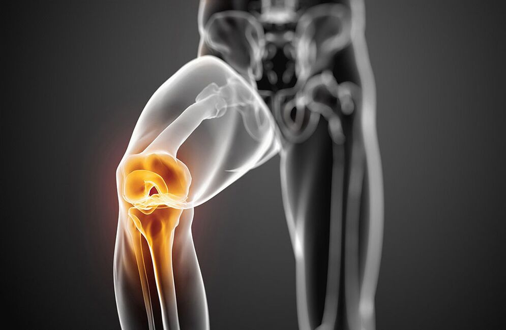
Arthrosis with damage to the knee joints - symptoms
The lesion of the knee joints with arthrosis is called gonarthrosis.Primary gonarthrosis develops in menopause women.The reasons for the secondary are the most frequent injuries to the knee joint and the disruption of the static with a curvature of the spine, flat legs.Patients complain of knee pain, which occurs during movements, especially when walking up the stairs.The pain is localized in front or inside the knee joint.The movements in the joint are limited: first bending and later expansion.When you move, a crunch often appears.With the development of reactive synovitis, the pain during movements intensifies and is worried at rest.Swelling of the joint, pain during palpation, redness (hyperemia) and fever of the skin are determined.Over time, due to bone growth, there is a deformity of the knee joints.
Arthrosis with damage to the hip joints - symptoms
The lesion of the hip joints is called coxarthrosis.This is the most severe form of arthrosis.The causes of the disease may be congenital dysplasia of the hip joints, injuries, menopause.Patients have joint pain during movements, upright.The restriction of movements in the joint gradually increases (first internal and external rotation, later flexion).There is a lame related to the shortening of the limb.With bilateral damage, the gait is typical.Atrophy of the muscles of the thighs and ass develops.There is no swelling of the joints with catrose.Palpation determines limited pain in the head of the femur.
In the initial stage of arthrosis, joint functions are preserved.With the more increasing development of the disease, it is first temporarily restricted and then the ability to work is completely lost, the patient loses the ability of self -care, needs external help.
The causes of arthrosis
Arthrosis is based on the primary degeneration of the articular cartilage with the accompanying destructive changes in the bones that form the joint.Such degeneration occurs as a result of an imbalance between the mechanical loads on the surface of the cartilage joint and the possibility of compensation for this load.
In the development of degenerative changes in the articular cartilage, several factors can simultaneously participate:
- Functional overloads, including professional, household and sports, causing cartilage mycotraum;
- joint injuries;
- infectious and not -specific inflammation of the joint;
- Joint dysplasia, which leads to a disruption of the comparison of joint surfaces;
- Disruption of body static as a result of spinal curvature (kyphosis, scoliosis, pathological lordosis, etc.), flat legs;
- Chronic Hemarthrosis:
- diseases with metabolic disorders (gout, obesity, chondrocalcinosis);
- Osteodistrophy or Paja's disease;
- osteomyelitis;
- pathology of the peripheral nervous system with loss of sensitivity;
- Endocrine pathology (acromegaly, diabetes, amenorrhea, hyperthyroidism);
- Hereditary tendency.
The risk factors of arthrosis include adult age, female, obesity.
Development mechanism
Metabolic disorders in cartilage are based on quantitative and qualitative changes in the main substance of cartilage.The main substance consists of proteoglycans that provide stability of collagen.The development of arthrosis is accompanied by insufficient formation or increased destruction of cartilage components.
With osteoarthritis in cartilage tissue, the content of hyaluronic acid, chondroitin and keratin decreases.In addition, altered proteoglycans lose the ability to retain water.It is absorbed by collagen, which swells, leading to a decrease in cartilage resistance.
If chondrocytes are damaged, they begin to produce collagen and proteoglycans, which are not characteristic of normal cartilage tissue.These altered substances cause the loss of biochemical properties of cartilage.
Immune disorders are of great importance for the development of arthrosis.The destruction of cartilage proteoglycans is accompanied by the appearance of immune reactions by the cell and humoral type.In turn, this causes progressive fibrosis and sclerosis of the synovial membrane, pathological changes in the intra -articular synovial fluid and disorder of cartilage.The lower synovial sheath supports the progression of degenerative changes in joint cartilage.
The hereditary factor has a certain value in the development of arthrosis.
Classification of arthrosis
Arthrosis is divided into two groups: primary and secondary.
In distribution (primary arthrosis):
- local (with a fault of three joints)
- General or generalized polyarthrosis (damage to three joints or more).
Depending on the destination (secondary):
- A. Tazobida joint (Cokeartrosis);
- A. The knee joint (gonarthrosis);
- A. The elbow joint;
- A. The shoulder joint;
- A. Spine;
- A. Department of cervical department (non -coarthrosis);
- A. Hands;
- A. Ankle pair (cruising)
- A. Stop.
Through etiology:
- after -traumatic
- metabolic
- Due to endocrine pathology.
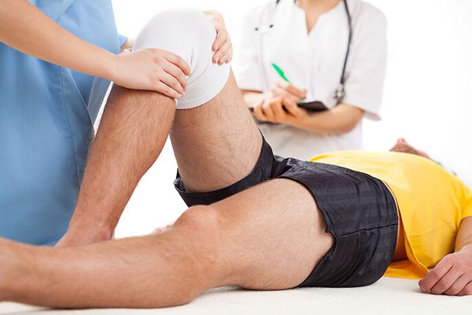
Diagnosis of arthrosis
The variety of clinical manifestations and variants of arthrosis impedes early diagnosis of the disease.The outbreak of the diagnosis is also associated with the lack of specific symptoms, the hidden onset of the disease.Of great importance is the definition of the factors contributing to the development of arthrosis:
- chronic joint trauma;
- long -term performance of stereotypical movements;
- physical activity of the joint for a certain time;
- impaired metabolism of salt or fat;
- Hereditary vices of the musculoskeletal system.
The X -Ray test is of the most important importance in the diagnosis of arthrosis.The radiography of the two knees is performed in a direct position, the bent position, in addition to the lateral position.The classic signs of arthrosis of radiography are: narrowing of the joint precipice, the presence of osteophytes, subchondral bone sclerosis and subchondral cysts.There are the following stages of radiological changes in arthrosis:
- 0 - No changes.
- I - radiologically suspicious signs.
- II - minimal changes (slight narrowing of the joint gap, subsidiary osteosclerosis, single osteophytes).
- III - moderate manifestations (moderate narrowing of the charter, multiple osteophytes).
- IV- Expressed changes (the joint difference is not visible, many coarse osteophytes are determined), often there is synovitis.
No additional tools are required in the presence of these symptoms.
In the absence or low weight, joints, MRI, scintigraphy are performed.
Clinical tests of blood, urine and intra -articular synovial fluid are not included in the list of mandatory studies to diagnose arthrosis.But these tests are necessary to exclude such joint pathologies.
The main clinical and diagnostic signs of arthrosis:
- Mechanical pain in the joints;
- fatigue;
- feeling of instability in the joints of the lower limbs;
- Damage to the joints of the first toe of the foot and arms;
- the gradual onset of the disease;
- slow progressive current;
- joint deformity;
- hypotrophy of the regional muscles;
- repetitive synovitis;
- restriction of joint movements;
- X -ray is changing.
Arthrosis should be differentiated with damage to the joints with rheumatoid arthritis, infectious, metabolic and reactive arthritis.
Rheumatoid arthritis, unlike arthrosis, begins with inflammation of the small joints of the hands and feet.It is characterized by intense pain from the inflammatory type, morning stiffness of the joints, the presence of rheumatoid nodes.
Gotric arthritis is mainly found in men.Characteristic is high local activity with acute paroxysmal pain in the first plus-phalangal joint of the thumb of the foot.With gout, the presence of tofu is typical that there is a "blow" on the X -ray.
Psoriatic arthritis is characterized by skin lesions, especially the scalp, spindle-form of deformity of the fingers and a bright raspberry color of the skin over the affected joints.
Infectious arthritis is characterized by acute start, rapid development and course, acute pain, fever and effectiveness of antibacterial therapy.
Arthrosis
The treatment of arthrosis should be long, complicated.The basic principles of treatment of arthrosis:
- The unloading of the joints (the right way of mobility and mechanical loads, dosed walking, weight loss, exclusion of prolonged standing, wearing weights, strengthening the musculoskeletal apparatus, using physiotherapy exercises, massage, electrical stimulation).
- Conservative correction of static disorders (use of orthopedic shoes, corsets, supervisory organs).
- The impact on general metabolism and blood circulation (the use of biostimulants, vasodilating drugs, balneotherapy and physiotherapy courses twice a year).
- Elimination of reactive synovitis, anti -inflammatory therapy.
Patients with arthrosis show a diet with a limitation of salt, sugar, strong tea, coffee, smoked meats, sharp dishes.This improves the sensitivity of vascular and joint receptors, restores the tone of blood vessels, normalizes metabolism in chondrocytes.With arthrosis, it is necessary to drink enough fluid (at least 8 glasses of water a day).
The treatment of arthrosis drug involves the use of rapid activity of anti -inflammatory and painkillers (non -steroidal anti -inflammatory drugs -NSAPs), basic drugs -hondroprotectors.No?Non-selective and selective TSO-2 inhibitors are used by NSAIDs.
NSAIDs in the form of ointment or gel are used as local therapy for the affected joints.
In the presence of reactive synovitis, tendonitis or tendovaginitis, when the treatment of NSAIDs is ineffective, appropriate intra -articular or intramuscular administration of corticosteroids.
Basic therapy with chondroprotectors (chondroitin, glucosamine, hyaluronic acid) is used to prevent degeneration of cartilage joints.
The treatment of chondroprotectors is shown in the clinical and radiological stages of arthrosis I-III.
In addition to direct chondroprotectors, medicines are used that stimulate the repair of cartilage tissue (biogenic stimulants).These drugs are used during remission in the absence of reactive synovitis.
Arthrosis also shows medicines that improve microcirculation.In the presence of varicose veins of the lower limbs, a correction of venous blood flow is required.
In patients with arthrosis, it is necessary to diagnose and treat osteoporosis in a timely manner.
Physiotherapy of arthrosis
Physical methods of treatment are also associated with the basic therapy of arthrosis.Under their influence, metabolic processes are stimulated, the microcirculation of the blood and the tissue fluid is restored to neurogmoral regulation.
The complex of treatment with arthrosis includes induction, microwave therapy, pulse currents, drug electrophoresis and magnetotherapy.To eliminate synovitis, ultraviolet irradiation of the area of the affected joints at erythema doses use an electric field with ultra high frequency, electrophoresis with analine, demexide or hydrocortisone.
To prevent the progression of arthrosis, it is recommended to reduce body weight, to avoid increased joint loads, walking in a suppressed area, increased humidity and hypothermia.Individual selection of shoes and supervisors is important.
Gonarthrosis shows regular exercise, swimming, cycling, strengthens the muscles.Classes of heavy and athletics, football is not recommended.
Therapeutic exercises are performed differently, in a sitting position lying in the pool.Movements should not be intense, traumatic, their volume and number of repetitions increase gradually, avoiding overload.
Popular and effective methods of treating arthrosis also include massage and kineotherapy.
With significant changes in the joints with deformity, it is recommended to limit mobility, surgical treatment.Arthroplasty, endoprothetics, osteotomy are performed.
The disease prognosis
Primary arthrosis rarely leads to complete damage.In the presence of reactive synovitis, patients temporarily become disabled and are sometimes forced to change the profession.In secondary cocarchia, the prognosis is less favorable due to the rapidly progressive course of the disease with the development of significant impaired joint functions.In such cases, damage can occur in several years of disease.
Prevention of arthrosis
Primary prevention of arthrosis should begin in childhood.This is as follows:
- prevention and treatment of scoliosis;
- Flat leg correction with the help of special leaders;
- Physical education classes to strengthen muscles and relationships;
- rational nutrition and prevention of metabolic disorders;
- limiting heavy sports in childhood and adolescence;
- variable work sitting on a table with walking;
- Proper labor organization and other employees in enterprises where there is great physical activity.
Secondary prophylaxis provides for measures that prevent the development of a recurrent reactive synovitis.These include dosed walking, restriction of physical exertion, walking with support and other measures that unload the joints.In severe symptoms of arthrosis, it is necessary to constantly take basic medicines.General strengthening therapy, improvement of blood circulation and metabolism, annual spa treatment is recommended.
Which doctor will go to?
- Rheumatologist
- Orthopedist